Oculoplastics
Oculoplastics, also known as oculoplastic surgery or ophthalmic plastic and reconstructive surgery, is a specialised branch of ophthalmology that deals with the surgical and non-surgical treatment of the eyelids, lacrimal (tear) system, orbit (eye socket), and surrounding facial structures.
Oculoplastic surgery is unique in that it combines elements of ophthalmology, plastic surgery, and facial reconstructive surgery to address both functional and cosmetic issues around the eyes. Oculoplastic surgeons are trained to perform procedures that improve both the function and appearance of these areas.
At Wollongong Eye Specialists, we can offer our patients expertise in this area, specifically for the procedures detailed below.
Please do not hesitate to reach out to us if you would like more information, seek a consultation, or would like to organise a referral to our care.
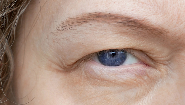
Ptosis (drooping eyelid)
Ptosis is the drooping of the upper eyelid, which can affect one or both eyes, and may be caused by muscle weakness, nerve damage, or other underlying conditions, potentially impairing vision.
Ptosis is treated based on its severity and cause, often with eyelid surgery, which is also referred to as ptosis repair, to tighten the muscles that lift the eyelid.
Blepharoplasty
Blepharoplasty is a surgical procedure that involves the removal or repositioning of excess skin, fat, and sometimes muscle from the upper and/or lower eyelids and is performed for both functional and cosmetic reasons.
Types of Blepharoplasty:
- Upper Blepharoplasty focuses on the upper eyelids and is performed to remove excess skin that may droop over the eyelashes, impairing vision. It can also improve the appearance of droopy eyelids.
- Lower Blepharoplasty focuses on the lower eyelids and is performed to remove bags under the eyes, reduce puffiness, and smooth out wrinkles or loose skin. It can also address fat prolapse, where fat around the eye pushes forward, creating a bulging appearance of the eye.
About the procedure:
The surgery is typically performed under local anaesthesia or general anaesthesia.
For upper blepharoplasty, an incision is made in the natural crease of the upper eyelid to remove or reposition excess skin, fat, and muscle. For lower blepharoplasty, the incision may be made just below the lower lash line or inside the eyelid to access and remove or reposition fat and tighten or remove loose skin.
The incisions are carefully closed with sutures, leaving minimal visible scars.
Patients may experience bruising, swelling, and mild discomfort for several days to weeks following the surgery. Sutures are typically removed within a week, and patients can usually return to normal activities within 1-2 weeks. Complete healing and results may take a few months as the tissues settle.
The benefits of blepharoplasty include improvement of visual function for those with drooping upper eyelids affecting vision and a more youthful and rested appearance, reducing the signs of ageing around the eyes.
Lid Lesions
A lid lesion refers to any abnormal growth or change in the skin of the eyelids. These lesions can vary widely in appearance and can be benign or malignant.
Types of Lid Lesions include:
Benign Lesions:
- Chalazion (blocked oil glands) or sebaceous cysts (filled with oil or keratin).
- Skin Tags which are small, soft growths
- Papillomas – benign tumours caused by the human papillomavirus often appearing as warty growths.
Malignant Lesions:
- Basal Cell Carcinoma (BCC): The most common type of skin cancer, usually appearing as a shiny bump or a flat, pinkish patch.
- Squamous Cell Carcinoma (SCC): A more aggressive form of skin cancer that can appear as a firm, red nodule or a flat lesion.
- Melanoma: A serious form of skin cancer that can appear as a dark mole or spot.
Treatment of Lid Lesions:
The treatment for lid lesions depends on the type, size, location, and whether they are benign or malignant. Here are common treatment approaches:
- Observation: For small, benign lesions that do not cause symptoms or cosmetic concerns, the doctor may recommend monitoring without immediate treatment.
- Medications: Topical treatments like creams or ointments may be used for certain benign lesions, or steroid injections can reduce inflammation or shrink cyst like lesions.
- Excision: The lesion is surgically removed, often under local anaesthesia. This is common for larger or suspicious lesions to ensure complete removal and to send samples for biopsy.
- Mohs Micrographic Surgery: A specialized technique for removing skin cancer while preserving as much healthy tissue as possible, particularly useful for basal cell or squamous cell carcinomas.
- Cryotherapy: Freezing the lesion with liquid nitrogen can be effective for certain benign growths like warts or precancerous lesions.
- Laser therapy: May be used to treat specific types of lesions, such as vascular lesions or for cosmetic improvement.
- Reconstruction: After excision, especially in the case of malignant lesions, reconstructive techniques may be necessary to restore the eyelid’s appearance and function. This can involve flap surgery or grafting.
Regular follow-up appointments may be required to monitor for any recurrence of lid lesions or to assess the healing process after treatment. Early detection and treatment are key to ensuring the best outcomes, especially for malignant lesions.
Dacryocystorhinostomy (DCR)
DCR is a surgical procedure with high success rates used to treat nasolacrimal duct obstruction which is when the tear drainage system is blocked, leading to excessive tearing, recurrent eye infections, and inflammation of the lacrimal sac which is the small pouch located in the inner corner of your eye, just above the tear duct.
The primary goal of DCR is to create a new pathway for tears to drain from the lacrimal sac into the nasal cavity. This restoration of normal tear drainage alleviates symptoms associated with tear duct obstruction.
There a two main types of DCR:
- External DCR which involves making a small incision on the side of the nose, near the inner corner of the eye and where the surgeon creates a new connection between the lacrimal sac and the nasal cavity. This technique allows for direct access to the anatomy and is often used in complex cases.
- Endoscopic DCR is a minimally invasive approach performed entirely through the nasal passages using an endoscope. No external incision is required, and the procedure is done through the nose.
About the procedure:
- The surgery is typically performed under local anaesthesia or general anaesthesia and during both external and endoscopic DCR, the surgeon creates an opening in the bone between the lacrimal sac and the nasal cavity.
- A silicone tube may be placed temporarily to maintain the new drainage pathway during the healing process which is usually removed after a few months.
Patients may experience mild discomfort, swelling, or bruising, particularly with external DCR and some may experience nasal congestion and minor nosebleeds. Most patients return to normal activities within a week or two, though full recovery may take longer.