Cornea
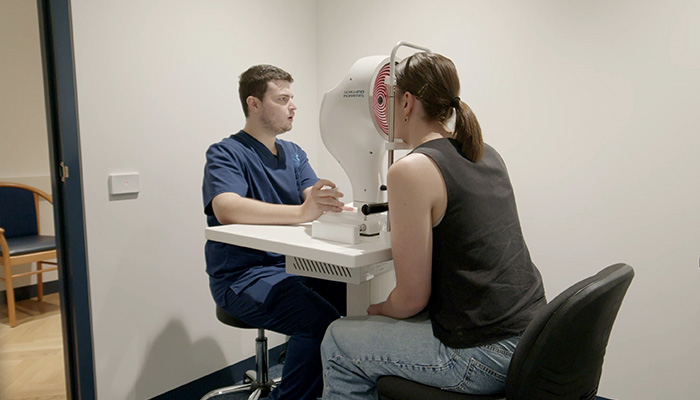
At Wollongong Eye Specialists, we can offer our patients expertise, and the regular monitoring and the management of the following conditions and diseases affecting the cornea, which is the clear, dome-shaped front surface of the eye.
Each of these conditions and their possible treatment and management strategies are detailed on this page.
Please do not hesitate to reach out to us if you would like more information, seek a consultation, or would like to organise a referral to our care for any of these ophthalmic issues.
Corneal Cross Linking (CXL)
Corneal Cross Linking (CXL) is designed to strengthen the cornea by promoting the bonding between collagen fibres within the cornea and is primarily used in the treatment of keratoconus, where the cornea becomes thin and cone-shaped, leading to distorted vision. CXL can significantly improve the stability of the cornea, reducing or halting the progression of keratoconus.
About the procedure:
The procedure typically begins with the removal of the epithelium (the outer layer of the cornea) to allow better absorption of a riboflavin solution used in the treatment.
A riboflavin solution is applied to the cornea for about 10-30 minutes depending on the protocol used. This substance helps to sensitively react with ultraviolet (UV) light.
After the riboflavin is applied, a specific wavelength of UV light is directed at the cornea for usually for around 10-30 minutes depending on the protocol used. This process activates the riboflavin and promotes the formation of new bonds between collagen fibres in the cornea.
After the procedure, a bandage contact lens may be placed on the eye to promote healing.
The procedure is day surgery and patients can return to the comfort of their own home to rest and recover.
About the recovery:
It usually takes 4-5 days for the epithelium to regenerate and a further 2-4 weeks before the vision in the eye approaches pre-operative levels. Antibiotic and anti-inflammatory eye drops are required for 6-8 weeks and appointments with your ophthalmologist are required and will be detailed during your consultation process.
It is usually necessary to be away from work for 7-10 days. Once the epithelium has healed, normal activities can commence and contact lenses can be worn again after two- three weeks but it may be four weeks before this can be done comfortably.
Descemet Membrane Endothelial Keratoplasty (DMEK)
DMEK is a specialised procedure, typically performed under local anaesthesia, used to treat conditions that affect the endothelium (the inner layer of the cornea), particularly Fuchs’ dystrophy and bullous keratopathy.
DMEK is primarily performed to replace the damaged endothelial layer of the cornea with healthy donor tissue which serves to improve vision and alleviate symptoms associated with corneal swelling and opacity.
About the procedure:
Beginning with the preparation of donor tissue, a thin layer of donor corneal tissue, specifically the Descemet membrane, along with the endothelial cells, is prepared.
A small incision is made in the cornea, and the diseased endothelial layer is removed. The donor tissue is then carefully inserted into the eye and positioned on the remaining stroma (the middle layer of the cornea). Air or fluid is often used to help the donor tissue adhere to the host cornea and ensure proper positioning. The incision is usually self-sealing, and stitches may not be necessary.
Benefits of DMEK and DSAEK:
This procedure generally results in a faster recovery time and improved visual outcomes compared to Penetrating Keratoplasty (PK), where the entire thickness of the cornea is replaced, and the procedure typically induces less astigmatism and maintains more of the cornea’s original structure.
About the recovery:
Patients usually experience improved vision within days to weeks after the surgery, although full recovery and stabilisation of vision may take several months. Regular follow-up appointments with your eye doctor are essential to monitor healing and ensure the donor tissue remains clear.
As with any surgical procedure, DMEK has potential risks including:
- Graft rejection.
- Detachment of the donor tissue.
- Infection
- Increased intraocular pressure which can lead to glaucoma
- Changes in vision due to irregular astigmatism.
DMEK is an effective option for patients with corneal endothelial dysfunction, both providing a minimally invasive way to restore vision while preserving the eye’s natural anatomy.
Descemet Stripping Automated Endothelial Keratoplasty (DSAEK)
Like DMEK, DSAEK is a procedure performed under local anaesthesia used to treat diseases of the corneal endothelium, primarily conditions like Fuchs’ endothelial dystrophy and bullous keratopathy.
This technique aims to replace the damaged endothelial layer of the cornea with healthy donor tissue to improve vision.
About the procedure:
Beginning with preparation of donor tissue, a thin layer of donor corneal tissue, specifically the Descemet membrane along with the endothelial cells, is prepared.
A small incision (usually around 2.5 to 3 mm) is made in the patient’s cornea, and the diseased endothelial layer is stripped away. The healthy donor tissue is then inserted into the eye, often rolled up to fit through the small incision. Once inside the eye, the donor tissue is unrolled and positioned over the remaining corneal stroma (the middle layer of the cornea). An air bubble may be introduced to help the donor tissue adhere properly to the host cornea. The incision is usually self-sealing, and sutures are often not needed.
Benefits DSAEK:
This procedure generally results in a faster recovery time and improved visual outcomes compared to Penetrating Keratoplasty (PK), where the entire thickness of the cornea is replaced, and the procedure typically induces less astigmatism and maintains more of the cornea’s original structure.
About the recovery:
Patients usually experience improved vision within days to weeks after the surgery, although full recovery and stabilisation of vision may take several months. Regular follow-up appointments with your eye doctor are essential to monitor healing and ensure the donor tissue remains clear.
As with any surgical procedure, DSAEK has potential risks including:
- Graft rejection.
- Detachment of the donor tissue.
- Infection
- Increased intraocular pressure which can lead to glaucoma
- Changes in vision due to irregular astigmatism.
DSAEK is an effective option for patients with corneal endothelial dysfunction, both providing a minimally invasive way to restore vision while preserving the eye’s natural anatomy.
Penetrating Keratoplasty (PK)
Penetrating Keratoplasty (PK), is a procedure performed under local or general anaesthesia used to replace all or a significant part of the cornea with healthy donor tissue and is typically performed to treat severe corneal diseases that affect vision, such as keratoconus, corneal scarring, Fuchs’ endothelial dystrophy, and other conditions that compromise the clarity or integrity of the cornea.
It is often indicated when other treatments, such as medications or less invasive surgeries, are insufficient.
About the procedure:
Beginning with the preparation of donor tissue, a full-thickness corneal graft is obtained from a donor.
A circular incision is made in the patient’s cornea to remove the diseased or damaged tissue. The donor cornea is then carefully positioned and sutured into place, these sutures can be adjusted post-operatively to achieve the desired curvature and visual outcome. This provides a new, clear surface for light to enter the eye, which can lead to better visual outcomes compared to the diseased cornea.
About the recovery:
Recovery from PK can be lengthy, typically taking several months to a year for full visual stabilisation. During this time patients may experience some discomfort, blurred vision, and sensitivity to light initially after the surgery.
Regular follow-up visits with your eye doctor are essential to monitor the healing process and manage any complications.
As with any major surgical procedure, PK carries risks, including:
- Graft rejection: The body may recognise the donor tissue as foreign and mount an immune response.
- Infection: There is a risk of postoperative infection that could threaten the graft.
- Glaucoma: Increased intraocular pressure may occur after surgery.
- Astigmatism: Changes in the shape of the cornea can lead to visual distortions.
- Corneal opacification or failure: The graft may not remain clear over time.
PK is a well-established surgical procedure for treating severe corneal diseases. While it can lead to significant improvements in vision, it requires careful patient selection and postoperative management to ensure best outcomes. Advances in corneal transplantation techniques, such as DSAEK and DMEK detailed earlier, have provided alternatives that may offer faster recovery and fewer complications, however PK remains an important option in the field of ophthalmology for certain cases.
Corneal Allogenic Intrastromal Ring Segment Transplant (CAIRS Transplant)
Corneal Allogenic Intrastromal Ring Segment Transplant (CAIRS Transplant) is a procedure that involves implanting a ring segment made from donor corneal tissue into the stroma (the middle layer) of the cornea. This technique is primarily used to treat keratoconus, where the cornea becomes thin and bulges outward, leading to distorted vision.
Its primary goal is to improve corneal shape and optical quality by providing additional support to the weakened cornea.
About the procedure:
Performed under local anaesthesia, a small incision is made in the cornea, and a pocket is created in the stromal layer where the ring segment will be placed. The allogenic ring segment is then carefully inserted into the created pocket, acting as a scaffold, providing mechanical support to the cornea. The incision may be sutured or left to heal naturally.
The benefits of CAIRS Transplant:
- CAIRS Transplant can help flatten the cornea, reducing the steepness associated with keratoconus.
- Improvements of visual acuity and reduction in the need for corrective lenses
- minimally invasive, it has a shorter recovery time compared to full corneal transplants
The recovery:
Patients may experience some discomfort or visual disturbances following the procedure, but these usually improve as healing progresses. Follow-up appointments are essential to monitor healing and assess visual outcomes. As with any procedure, CAIRS Transplant carries some risks, including:
- Infection
- Graft rejection (in the case of donor tissue)
- Corneal scarring
- Changes in vision.
- Potential for complications related to the ring segment itself
CAIRS Transplant is an innovative approach to managing keratoconus and other corneal conditions. By providing additional structural support to the cornea, this technique can lead to improved vision and quality of life for patients.